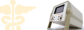
Light MD™ Therapy
 Trent Dilfer: Played professional football for 14 years, Super Bowl Champion with Baltimore Ravens.
![]() “In a sport like football it is critically important to treat injuries properly, and even more important to stay ahead of injuries. Without ART and Dr. Joe Leahy, I would not have been able to play as long or effectively as I did. It dramatically reduces pain, and keeps performance up. And now with the addition of Light MD therapy, ART works faster, and more completely. I went in to see Dr. Leahy in a lot of pain and after 20 minutes of APL and then ART, I had complete range of motion, and no pain, ready to resume workouts.”
“In a sport like football it is critically important to treat injuries properly, and even more important to stay ahead of injuries. Without ART and Dr. Joe Leahy, I would not have been able to play as long or effectively as I did. It dramatically reduces pain, and keeps performance up. And now with the addition of Light MD therapy, ART works faster, and more completely. I went in to see Dr. Leahy in a lot of pain and after 20 minutes of APL and then ART, I had complete range of motion, and no pain, ready to resume workouts.”
Amy Anaya: National Champion Weightlifter, training for 2012 Olympics
“I would not even be considering the Olympics if it were not for Dr. Leahy. His adjustments and ART are what keep me going, and helped get me past an injury.”
“I have never found a supplement to my chiropractic care that has worked as quickly and effectively as Light MD’s APLightsource 2000™. As a competitive athlete, being in the gym every day and training at the highest intensity possible is very important. APL has been an absolute blessing in healing weekly aches and pains in my wrist, knees, and hip. It is a fantastic device that I can’t imagine being without.”
Light MD (APL) therapy can have a remarkable effect on all areas of your body. It has been scientifically proven to be extremely effective at treating injuries, sprains/strains, inflammation, wounds, infections, cellulite, flaccid skin, scarring, stretch marks, and age or sun spots. And the list goes on. Until now, there has been no single method to treat all of these problems, until now…A Light MD (APL) is that treatment method.
APL will stimulate the release of ATP, the energy molecule of the cell, which allows cells to repair faster. APL stimulates the production of collagen, the essential protein used to repair and replace damaged, skin, ligaments, cartilage and tendons, and decrease scar tissue. APL will increase the vascularity of tissue. Tests conducted with the scanning laser Doppler demonstrate increases in microcirculation from 400% to 3200% after one treatment. Basically, it makes any tissue heal faster, studies show150-200% faster, and markedly stronger!
 Reduces Pain, Speeds Injury Recovery, Reduces Inflammation, Increases Cellular energy, Increases Tissue Strength, Increases Circulation. Here are some of the research abstracts that show what light therapy in general can do. What I have found after 2 years of personal experience with most of the available lasers, and LED units, Light MD is literally “light years ahead” of the competitions.
1. Lasers Surg Med. 2010 Aug;42(6):553-8.
Anti-inflammatory effects of low-level light emitting diode therapy on Achillestendinitis in rats.
Xavier M, David DR, de Souza RA, Arrieiro AN, Miranda H, Santana ET, Silva JA Jr,Salgado MA, Aimbire F, Albertini R.
Institute of Research and Development, IP&D, Vale do Paraiba University, UNIVAP, Av. Shishima Hifumi, 2911, 12244-000 São José dos Campos, São Paulo, Brazil.
BACKGROUND AND OBJECTIVES: The present study investigated the effects oflow-level light emitting diode (LED) therapy (880 +/- 10 nm) on inflammatoryprocess in a experimental model of Achilles tendinitis induced by collagenase.STUDY DESIGN/MATERIALS AND METHODS: Fifty-six male Wistar were separated intoseven groups (n = 8), three groups in the experimental period of 7 days and four groups in the experimental period of 14 days, the control group (CONT),tendinitis group (TEND), LED therapy group (LEDT) for both experimental periods, and LED therapy group 7th to 14th day (LEDT delay) for 14 days experimental period. The LED parameters was 22 mW CW of optical output power, distributed inan irradiation area of 0.5 cm(2), with an irradiation time of 170 seconds, the applied energy density was 7.5 J/cm(2) in contact. The therapy was initiated 12hours after the tendinitis induction, with a 48-hour interval between their radiations. The histological analysis and inflammatory mediators were quantified.RESULTS: Our results showed that LED decreases the inflammatory cells influx and mRNA expression to IL-1 beta, IL-6, tumor necrosis factor-alpha (TNF-alpha) in both phase, and cyclooxygenase-2 (COX-2) just in initial phase (P < 0.05).
CONCLUSION: Our results suggest that the anti-inflammatory therapy with low-powerLED (880 nm) enhanced the tissue response in all groups. We can conclude that the LED was able to reduce signs of inflammation in collagenase-induced tendinitis inrats by reducing the number of inflammatory cells and decrease mRNA expression ofcytokines.
PMID: 20662032 [PubMed – indexed for MEDLINE]
Varying ratios of wavelengths in dual wavelength LED photomodulation alters gene expression profiles in human skin fibroblasts.
McDaniel DH, Weiss RA, Geronemus RG, Mazur C, Wilson S, Weiss MA.
Laser Skin & Vein Center of Virginia, Institute of Anti-Aging Research; Virginia Beach, Virginia 23462, USA. mail@lsvcv.com
BACKGROUND AND OBJECTIVE: LED photomodulation has been shown to profoundlyinfluence cellular behavior. A variety of parameters with LED photomodulation canalter cellular response in vitro. The effects of one visible and one infraredwavelength were evaluated to determine the optimal ratio to produce a netincrease in dermal collagen by altering the ratio of total energy output of each wavelength. The ratio between the two wavelengths (590 and 870 nm) was shifted in25% increments.STUDY DESIGN/MATERIALS AND METHODS: Human skin fibroblasts in culture were exposed to a 590/870 nm LED array with total combined energy density fixed at 4.0mW/cm.. The ratio of 590/870 nm tested parameters were: 100/0%, 75/25%, 50/50%,25/75%, and 0/100%. These ratios were delivered using pulsed duty cycle ofexposure (250 milliseconds “on” time/100 milliseconds “off” time/100 pulses) for a total energy fluence of 0.1 J/cm.. Gene expression was examined using commercially available extra cellular matrix and adhesion molecule RT PCR Arrays (SA Biosciences, Frederick, MD) at 24 hours post-exposure.RESULTS: Different expression profiles were noticed for each of the ratiosstudied. Overall, there was an average (in an 80 gene array) of 6% expression difference in up or down regulation between the arrays. The greatest increase in collagen I and decrease in collagenase (MMP-1) was observed with 75/25% ratio of 590/870 nm. The addition of increasing proportions of IR wavelengths causes alteration in gene expression profile. The ratios of the wavelengths causedvariation in magnitude of expression.CONCLUSIONS: Cell metabolism and gene expression can be altered by simultaneous exposure to multiple wavelengths of low energy light. Varying the ratios of specific wavelength intensity in both visible and near infrared light therapy canstrongly influence resulting fibroblast gene expression patterns.
PMID: 20662030 [PubMed – indexed for MEDLINE]
1. Lasers Surg Med. 2010 Aug;42(6):597-601.
Prophylactic low-level light therapy for the treatment of hypertrophic scars and keloids: a case series.
Barolet D, Boucher A.
BACKGROUND AND OBJECTIVES: Hypertrophic and keloid scars result from alterations in the wound healing process. Treating abnormal scars remains an important challenge. The aim of this case series was to investigate the effectiveness ofnear infrared (NIR) light emitting diode (LED) treatment as a prophylactic method to alter the wound healing process in order to avoid or attenuate the formation of hypertrophic scars or keloids.STUDY DESIGN/PATIENTS AND METHODS: Three patients (age 27-57) of phototypes I-IIIwith hypertrophic scars or keloids due to acne or surgery participated in thiscase series. Following scar revision by surgery or CO(2) laser ablation onbilateral areas, one scar was treated daily by the patient at home withnon-thermal, non-ablative NIR LED (805 nm at 30 mW/cm(2)) for 30 days. Efficacyassessments, conducted up to a year post-treatment, included the Vancouver Scarscale (VSS), clinical global assessment of digital photographs, and quantitative profilometry analysis using PRIMOS. Safety was documented by adverse effectsmonitoring.RESULTS: Significant improvements on the NIR-treated versus the control scar were seen in all efficacy measures. No significant treatment-related adverse effects were reported.CONCLUSION: Possible mechanisms involved are inhibition of TGF-beta I expression.Further studies in larger group of patients are needed to evaluate this promising technique.
PMID: 20662038 [PubMed – indexed for MEDLINE]
1. Photodiagnosis Photodyn Ther. 2010 Mar;7(1):44-9. Epub 2010 Jan 15.
Effect of light emitting diodes in the photodynamic therapy of rheumatoidarthritis.
Neupane J, Ghimire S, Shakya S, Chaudhary L, Shrivastava VP.
College of Biomedical Engineering and Applied Sciences, Dhana Ganesh, HandigaunMarg, Kathmandu, Nepal. jiteshneupane@gmail.com
BACKGROUND: Complex and painful surgical removal of synovium was replaced byarthroscopic synovectomy as an early treatment of rheumatoid arthritis (RA),which being limited to bigger joints, was replaced by laser synovectomy. Having been more time consuming, laser photodynamic therapy (PDT) replaced this method. Due to thermal side effects of laser PDT, an alternative source of light has been sought. Therefore, to make RA treatment cheaper, less hazardous and suitable according to anatomical geometry, light emitting diodes (LEDs) were used in this study as a potential source of light.METHODS: Red, white, yellow and infra-red (IR) LEDs were tested to measure the optical penetration for soft tissue and their scattering. In vitro study of the cellular response of normal and inflamed lymphocytes from healthy and RA patients was conducted respectively. Methotrexate was injected as photosensitizer toachieve cell-specific precision.RESULTS: IR LEDs showed the maximum penetration and least scattering of all LEDs used. Specimen with drug administration and with subsequent exposure to IR LEDs exhibited massive suppression of inflamed activated lymphocytes in comparison to other controls.CONCLUSION: The properly selected wavelength and intensity of light beam were incident with great precision so that they would not affect unwanted cells, but inflamed activated cells were suppressed due to intense light energy following Methotrexate injection. Without invasion, IR LED PDT showed an effective and cheaper treatment solution for RA.